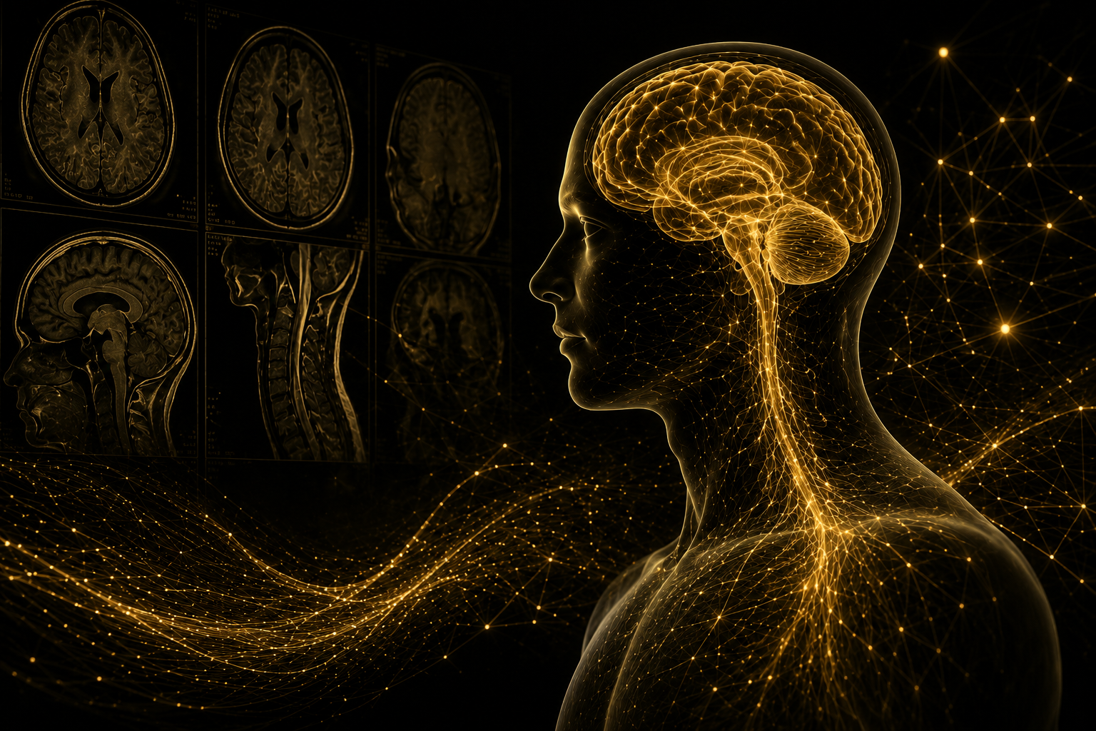
Why Imaging May Appear Normal Despite Persistent Symptoms
Understanding Why Normal CT or MRI Findings Do Not Always Mean Normal Neurologic or Autonomic Function
Persistent symptoms can occur even when CT or MRI studies do not identify a major structural abnormality. This is particularly common in conditions involving concussion, dysautonomia, POTS, exercise intolerance, post-viral illness, and other disorders of physiologic regulation.
CT and MRI are essential for identifying structural problems such as bleeding, fractures, tumors, stroke, swelling, and significant tissue injury. However, many persistent symptoms arise from abnormalities in how the nervous system regulates blood flow, heart rate, blood pressure, balance, vision, cognition, exercise response, and recovery.
These physiologic disturbances may be clinically significant even when routine structural imaging appears normal.
Structural Pathology vs Functional Pathology
MRI and CT imaging are extremely important tools in modern medicine. They are highly effective for identifying serious structural brain abnormalities such as bleeding, fracture, stroke, tumor, severe spinal cord compression, or other significant tissue injury.
These studies are often critical for ruling out dangerous or life-threatening conditions. However, many concussion-related and autonomic symptoms involve physiologic dysfunction (how the brain works) rather than large structural abnormalities visible on standard imaging studies. In these situations, even when imaging does not identify pathology the nervous system may still not be regulating, communicating, or recovering normally despite the absence of obvious findings on MRI or CT imaging.
Examples may include dysfunction involving:
Autonomic nervous system regulation
Neurocardiac communication pathways
Blood flow regulation
Exercise tolerance and physiologic recovery
Vestibular and balance systems
Oculomotor control and visual processing
Sensory overstimulation
Cognitive endurance and fatigue regulation
As a result, many patients experience significant symptoms despite having imaging studies that appear “normal.” The lack of significant imaging findings does not mean the symptoms are psychological or insignificant. Symptoms in the presence of normal imaging may reflect dysfunction occurring at the physiologic, microscopic, metabolic, or brain network-based levels that are often not seen through conventional structural imaging, like CT and MRI.
Understanding the difference between structural injury and functional physiologic dysfunction is an important part of evaluating persistent symptoms following concussion, dysautonomia, autonomic dysfunction, or other neurologic stressors.

Concussion Is Defined as a Functional Brain Injury
Concussion most frequently involves disturbance of brain function rather than obvious structural injury visible on routine imaging studies. If the imaging is abnormal the patient may still have concussion with identified pathology. Following concussion, the brain experiences “neurometabolic” changes. These changes involve brain blood control, energy regulation, chemical changes, autonomic nervous system activity, sensory processing, exercise tolerance, and physiologic recovery.
These functional disturbances can contribute to symptoms such as:
Brain fog
Head pressure or headaches
Dizziness and motion sensitivity
Fatigue and reduced endurance
Exercise intolerance
Difficulty concentrating
Visual sensitivity
Sleep disruption
Heart rate variability or autonomic instability
Delayed recovery following physical or cognitive activity
In many patients, these symptoms reflect disruption in how neurologic systems regulate and communicate rather than major tissue destruction visible on MRI or CT imaging. Concussive injury is not able to be identified on readily accessible advanced imaging like CT and MRI.
Concussion may also affect the autonomic nervous system, the system responsible for regulating automatic functions such as heart rate, blood pressure, circulation, temperature regulation, and physiologic recovery. When these systems become dysregulated, patients may develop symptoms involving upright intolerance, fatigue, exercise intolerance, dizziness, or physiologic overstimulation despite relatively normal structural imaging findings.
This is one reason concussion recovery can sometimes become prolonged or more complex than expected. Symptoms may fluctuate significantly depending upon cognitive load, sleep quality, stress exposure, environmental stimulation, hydration status, exercise demands, and autonomic recovery capacity.
At NeuroSport, evaluation focuses not only on whether imaging appears normal, but also on how the brain and body are functioning together during recovery.
For a more detailed concussion-specific explanation, read Why Is My MRI Normal After a Concussion? That page explains what routine CT and MRI studies are designed to detect and why concussion symptoms may persist despite normal structural imaging.
Why Dysautonomia, POTS, and Autonomic Dysfunction May Not Appear on Standard Imaging
Dysautonomia refers to dysfunction of the autonomic nervous system, the system responsible for regulating automatic body functions such as heart rate, blood pressure, circulation, digestion, temperature regulation, and physiologic recovery.
These disorders may involve impaired cardiovascular regulation, blood-volume distribution, cerebral blood flow, vascular tone, and recovery physiology rather than a visible structural lesion.
Unlike conditions involving major structural injury, many autonomic disorders primarily involve abnormalities in regulation and communication rather than obvious tissue damage visible on MRI or CT imaging. For example, patients with dysautonomia or POTS may experience the following symptoms even when structural imaging studies appear normal:
Orthostatic intolerance
Blood pooling
Rapid heart rate upon standing
Heat intolerance
Exercise intolerance
Fatigue and reduced physiologic reserve
Adrenaline surges or physiologic overstimulation
Dizziness or near-fainting episodes
Delayed recovery after activity
This occurs because autonomic dysfunction often involves abnormalities in how physiologic systems regulate circulation, cardiovascular response, blood vessel tone, recovery mechanisms, and communication between the brain, heart, and vascular system.
In many cases, dysfunction may occur at physiologic, microscopic, metabolic, or network-based levels that are not fully visualized on standard structural imaging studies. This does not mean symptoms are minor or insignificant. Autonomic dysfunction can substantially affect exercise tolerance, cognitive endurance, work capacity, school participation, environmental tolerance, and daily functioning despite the absence of major imaging abnormalities.
Understanding this distinction is important when evaluating persistent symptoms following concussion, viral illness, physiologic stress, or other neurologic and autonomic conditions.
The Frustration of Persisting Symptoms Despite “Normal” Testing
Patients understandably become frustrated when symptoms persist despite normal MRI, CT, laboratory, or cardiac testing. They may begin questioning whether their symptoms are real or whether something important has been missed.
Normal structural testing is reassuring, but it does not measure every aspect of neurologic and autonomic function. Physiologic dysfunction may involve blood-flow regulation, exercise response, sensory processing, balance, cognition, sleep, or autonomic control.
In concussion and autonomic disorders, symptoms may arise from disturbances from different domains involving:
Blood flow regulation within the brain and body
Exercise intolerance
Autonomic nervous system activity
Sensory processing
Vestibular function
Neurocardiac communication
Sleep regulation
Cognitive endurance and fatigue management
These very important physiologic systems can become dysregulated even when conventional imaging studies do not reveal major structural abnormalities. Another challenge is that symptoms often fluctuate. Patients may experience periods of relative stability followed by symptom flares triggered by heat, stress, illness, poor sleep, cognitive overload, environmental stimulation, travel, or physical overexertion. This variability can make symptoms appear inconsistent despite an underlying physiologic basis.
Additionally, many patients gradually adapt their activities over time. Individuals may reduce exercise, avoid symptom-provoking situations, sit more frequently, pace their activities, or unconsciously limit daily demands in ways that partially mask the severity of dysfunction.
At NeuroSport, evaluation focuses on understanding how physiologic systems are functioning together rather than relying solely on whether imaging appears normal or abnormal. The goal is to better identify the patterns contributing to persistent symptoms, exercise intolerance, autonomic instability, and reduced recovery capacity.
Objective Evaluation Still Matters
Although standard imaging studies may appear normal, objective evaluation remains extremely important when assessing persistent concussion-related or autonomic symptoms.
At NeuroSport, evaluation focuses on identifying measurable physiologic patterns that may help explain symptom persistence, exercise intolerance, autonomic instability, dizziness, cognitive fatigue, or delayed recovery. Depending upon the clinical presentation, assessment may include:
Orthostatic vital sign analysis
NASA Lean Testing
Exercise tolerance assessment
Heart rate and physiologic response monitoring
Autonomic symptom pattern analysis
Oculomotor and vestibular evaluation
Cervical spine assessment
Cognitive screening and processing evaluation
Activity tolerance analysis
Recovery pattern assessment
The goal is not simply to determine whether imaging is normal or abnormal, but to better understand how the nervous system and body are functioning together under physiologic demand. This systems-based approach may help identify patterns that are not always visible through structural imaging alone, particularly in patients with concussion, dysautonomia, exercise intolerance, or persistent neurologic symptoms.
At NeuroSport, emphasis is placed on integrating patient history, symptom patterns, physiologic response, functional capacity, and objective findings to guide more individualized evaluation and recovery planning.
A Normal MRI Does Not Always Mean Full Physiologic Recovery
A normal MRI or CT scan is reassuring because it helps rule out dangerous structural injury. However, normal imaging does not necessarily mean that all neurologic, autonomic, or physiologic systems have fully recovered.
Many patients with persistent concussion-related or autonomic symptoms continue to experience measurable limitations involving:
Exercise tolerance
Cognitive endurance
Upright tolerance
Heart rate and blood pressure regulation
Balance and dizziness
Sensory processing
Fatigue management
Recovery stability following activity
In some cases, patients may appear outwardly healthy while still struggling with significant physiologic limitations during school, work, athletics, travel, or daily activities.
This distinction is particularly important in conditions involving concussion, dysautonomia, autonomic dysfunction, post-viral illness, or prolonged physiologic stress. Functional recovery may lag behind structural recovery, especially when autonomic regulation, exercise tolerance, or recovery systems remain disrupted.
Understanding the difference between “normal imaging” and “normal physiologic function” is an important part of evaluating persistent symptoms and developing more individualized recovery strategies.
The NeuroSport Approach
The NeuroSport evaluation is designed to look beyond whether imaging appears “normal” or “abnormal” and instead focus on how physiologic systems are functioning together.
Many persistent concussion and autonomic presentations involve overlapping factors affecting exercise tolerance, autonomic regulation, cognitive endurance, vestibular function, recovery physiology, cervical spine mechanics, and activity tolerance. Understanding these interactions is often important when symptoms persist despite relatively normal structural imaging findings.
Our evidence guided evaluations emphasize a systems-based approach that integrates patient history, symptom patterns, physiologic response, objective testing, and functional performance rather than relying on imaging findings alone. Patients with concussion-related and/or autonomic dysfunction can prove their injury through a through examination process.
The goal is not simply to identify structural injury, but to better understand the physiologic contributors affecting recovery, daily function, exercise capacity, and symptom persistence.
Normal Imaging Does Not End the Evaluation
Persistent dizziness, fatigue, brain fog, exercise intolerance, rapid heart rate, balance problems, or cognitive difficulty may reflect dysfunction that routine structural imaging does not measure.
NeuroSport evaluates concussion, post-concussion syndrome, dysautonomia, POTS, autonomic dysfunction, and exercise intolerance using a systems-based clinical approach. The goal is to identify measurable physiologic contributors and develop an individualized plan for recovery.